Interior Protein Network Diagram Of Cell Membrane
Free Printable Interior Protein Network Diagram Of Cell Membrane

Some cells use extrusion in which water is ejected through contractile vacuoles.
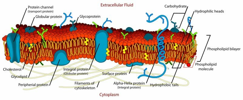
Interior protein network diagram of cell membrane. Every cell membrane is composed of phospholipids in a bilayer. The two major classes of proteins in the cell membrane are integral proteins a protein that spans the lipids bilayer of membranes which span the hydrophobic interior of the bilayer and peripheral proteins a protein that is more loosely associated with the membrane surface which are more loosely associated with the surface of the lipid. Phospholipid bilayer transmembrane proteins interior protein network cell surface markers. An important feature of the membrane is that it remains fluid.
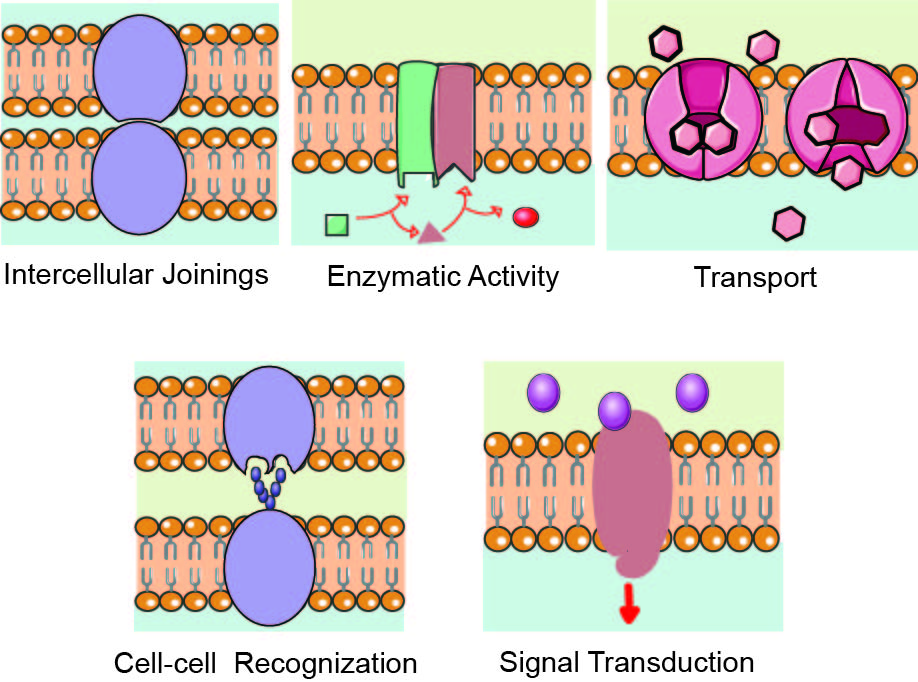
Such an assumption may also explain the impact that flotillins have on the development of cell wall. The cell membrane also known as the plasma membrane pm or cytoplasmic membrane and historically referred to as the plasmalemma is a biological membrane that separates the interior of all cells from the outside environment the extracellular space which protects the cell from its environment. A protein on a cell wall that binds with specific molecules so that they can be absorbed into the cell. The plasma membrane carries markers that allow cells to recognize one another and can transmit signals to other cells via receptors.
The lipids and proteins in the cell membrane are not rigidly locked in place. The cell membrane has many proteins as well as other lipids such as cholesterol that are associated with the phospholipid bilayer. The semipermeable barrier that surrounds the cytoplasm of a cell. The cell membrane consists of a lipid bilayer including cholesterols a lipid component that.
Additionally the protein machinery that produces the cell wall moves. Requires expenditure of energy atp. Isosmotic regulation involves keeping cells isotonic with their environment. The other components of the membrane are embedded within the bilayer which provides a flexible matrix and at the same time.
The sars cov 2 is part of the coronavirus family with characteristic spike proteins that stud the surface and mediate viral recognition by the host cell ace2 receptor membrane fusion and entry. The currently accepted model of cell membrane structure which envisions the membrane as a mosaic of protein molecules drifting laterally in a fluid bilayer of phospholipids. Peripheral or intracellular membrane proteins spectrins and clathrins. B sheets folding back and forh in a cylinder around the membrane call a b barrel.


